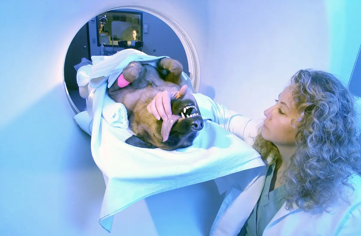
If your dog's MRI report mentions "intervertebral disc disease", "IVDD", a "disc extrusion", or a "disc protrusion", it is natural to feel shaken, especially if your dog is in pain or suddenly wobbly on its back legs. The reassuring truth is that many dogs with IVDD recover well, and the MRI you are reading is exactly the tool that helps your vet choose the right plan. Here is what these terms mean, how the two main types differ, how severity is graded, the one red flag that genuinely cannot wait, and what treatment usually involves.
What intervertebral disc disease actually is
Between each pair of bones in your dog's spine sits a small cushion called an intervertebral disc. It has a tough outer ring and a softer center, and it works as a shock absorber. In IVDD, a disc degenerates and either bursts or bulges, pushing material against the spinal cord that runs just above it. Because the spinal cord is what carries movement and sensation to the legs and tail, even a small amount of pressure in the wrong place can cause pain, weakness, or paralysis.
On MRI, a radiologist or neurologist looks at exactly where the disc material has gone, how much of the spinal cord is being squeezed, and whether the cord itself looks bruised or swollen. Those details are what turn a worrying word on a report into an actual plan.
Hansen type I versus type II — two different problems
Vets usually sort IVDD into two patterns, named after the researcher who first described them. They behave differently and tend to affect different dogs.
- Hansen type I (extrusion) is the sudden, forceful kind. The disc's center hardens and then bursts through the outer ring, firing material against the cord. It is the classic pattern in chondrodystrophic breeds — the short-legged, long-backed dogs such as Dachshunds, French Bulldogs, Beagles, and Shih Tzus — often in early to middle age. Signs can come on over hours.
- Hansen type II (protrusion) is the slow, chronic kind. The outer ring thickens and bulges gradually into the cord over months. It is more typical of older, larger dogs like German Shepherds and Labradors, and the signs tend to creep in rather than strike suddenly.
Knowing which pattern is present helps set expectations, because a sudden extrusion and a slow protrusion often call for different approaches and carry different outlooks.
Why MRI is the test of choice
A plain X-ray shows the bones of the spine, and it can hint that a disc space looks narrowed, but it cannot show the spinal cord or the disc material pressing on it. That is why an X-ray alone usually cannot tell your vet exactly which disc is the culprit or how badly the cord is affected. MRI, by contrast, shows the soft tissues directly and is considered the gold standard for localizing the compressed segment before any surgery. CT, sometimes combined with myelography (a dye study), is a very good alternative when MRI is not available or when a fast answer is needed out of hours.
Getting the location exactly right matters enormously, because a surgeon operating on the spine needs to know precisely which segment to decompress. The imaging is always read alongside your dog's neurological exam — the scan shows the anatomy, and the exam shows how the cord is actually working.
How vets grade the severity
Much of what happens next depends less on the MRI picture and more on how your dog is functioning. Vets describe severity along a well-recognized ladder, roughly from mildest to most serious:
- Painful but still walking normally
- Walking but wobbly and weak in the back legs (called paraparesis)
- Unable to walk but still able to move the legs
- Paralysed in the back legs, unable to move them at all
- Paralysed and no longer able to feel a firm pinch of the toes (loss of deep pain)
Where your dog sits on this ladder, and whether it is moving up or down it, is often more important for decision-making than the size of the disc bulge on the scan. This is exactly why imaging findings are always correlated with the hands-on exam by the treating vet.
The one red flag that can't wait
Please read this section even if you skip the rest. The single most important sign in IVDD is loss of deep pain perception. Deep pain is the deepest, most basic sensation in the leg; a vet tests it by firmly pinching a toe and watching for a true conscious reaction — the dog turning to look, whimpering, or trying to bite — not just a reflex pull-back. When a paralysed dog can no longer feel that firm pinch, the spinal cord injury is severe, and the clock starts to matter in a real way.
In this specific situation, the chance of walking again falls the longer deep pain stays absent, and surgery within roughly the first day or two gives the best odds. If your dog is suddenly paralysed in the hind end, do not wait to see whether it improves overnight — this is the classic scenario where hours count, and an emergency or specialist visit is warranted straight away.
Treatment — crate rest versus surgery
Broadly, there are two paths, and the right one depends on how severe and how sudden the signs are.
Conservative (non-surgical) management suits milder cases — a dog that is painful, or wobbly but still walking. It centers on strict crate rest for several weeks (genuinely strict — the point is to let the disc scar down without more movement), pain and anti-inflammatory medication, and a gradual, supervised return to activity. Many of these dogs do well, though rest really does have to mean rest.
Decompressive surgery, most often a hemilaminectomy, removes a small window of bone to lift the pressure off the spinal cord and clear out the disc material. It is generally recommended for dogs that are paralysed, that keep getting worse despite rest, that are in pain that cannot be controlled, or that have lost deep pain. For those dogs, surgery — and doing it promptly — usually offers the best chance of recovery.
What recovery usually looks like
Outlook depends heavily on that severity grade. Dogs that are still walking, and dogs that are down but still have deep pain, have a good chance of recovering with the appropriate treatment, though it can take weeks of rest and rehabilitation. The picture is more guarded once deep pain has been lost, and prompt surgery becomes the main lever on the outcome. Recovery is rarely instant either way; nursing care, physiotherapy, and patience all play a part, and your vet or a veterinary neurologist can give you a realistic picture for your individual dog. IVDD is also a spinal problem rather than a limb one, which distinguishes it from orthopedic causes of hind-leg trouble such as a cranial cruciate ligament tear or hip dysplasia that your vet may also consider.
Why a second read can help
A spinal MRI holds a lot of detail: which disc, which side, how compressed the cord looks, and whether the cord itself appears damaged — and those details steer the choice between rest and surgery, and how urgently. When an operation is on the table, having another set of expert eyes on the images can add real confidence to a hard decision. DocOrbit offers a veterinary second opinion on your animal's imaging that you can share with your own vet, which can be reassuring when you are weighing surgery against watchful waiting for a beloved pet.
Is IVDD in dogs an emergency?
It can be. A dog that is painful but still walking is usually not a same-day emergency, and many improve with rest and medication. But a dog that suddenly cannot use its back legs, and especially one that has lost the ability to feel a hard pinch of the toes, is a genuine emergency where hours matter. If your dog goes down quickly or drags its hind end, treat it as urgent and call a vet right away.
Can a dog recover from IVDD without surgery?
Yes, many dogs with milder IVDD recover well with conservative management: strict crate rest for several weeks, pain and anti-inflammatory medication, and a slow return to activity. This works best when the dog is still walking and only mildly wobbly. Dogs that are paralysed, that are getting worse, or that have lost deep pain generally do better with surgery, and time is important in those cases.
What does loss of deep pain mean in a dog?
Deep pain perception is the deepest, most basic sensation in the limb. A vet tests it by firmly pinching a toe and watching for a conscious reaction, such as turning to look or crying out, not just a reflex twitch. Losing it means the spinal cord injury is severe. It is the single most important sign on the exam, because the chance of walking again drops the longer it is absent, which is why it is treated as a surgical emergency.
Which dog breeds are most prone to IVDD?
The sudden, forceful disc extrusions of Hansen type I are classic in chondrodystrophic (short-legged, long-backed) breeds such as Dachshunds, French Bulldogs, Beagles, and Shih Tzus, often in early to middle age. The slower Hansen type II protrusions are more typical of older, larger breeds like German Shepherds and Labradors. Any dog can be affected, but breed and body type shift the odds.
Why is MRI used for IVDD instead of an X-ray?
A plain X-ray shows the bones but not the spinal cord or the disc material pressing on it, so it can suggest a problem but cannot confirm exactly where or how severe it is. MRI shows the soft tissues directly and is the gold standard for pinpointing which segment of the cord is compressed before surgery. CT, sometimes with myelography, is a good alternative when MRI is not available or a fast answer is needed.
Key takeaways
- IVDD is a disc pressing on the spinal cord; MRI is the gold standard for showing exactly where and how badly
- Hansen type I is the sudden extrusion of long-backed breeds; type II is the slow protrusion of older, larger dogs
- Severity is graded by function, from painful-but-walking through to paralysis and loss of deep pain
- Loss of deep pain is a surgical emergency where hours matter for the chance of walking again
- Milder cases often recover with strict crate rest and medication; more severe cases usually need decompressive surgery
Can I ask a veterinary radiologist about my pet's MRI report online?
Yes. Online veterinary radiology second opinions run entirely from home: you upload your images, with no CD to post and no second trip, and a subspecialist veterinary radiologist re-reads the study and writes a structured report you can give to your own vet. DocOrbit returns that signed second read in 24 to 48 hours. You can then message the veterinary radiologist with questions about the MRI and about what the report actually means for your pet.
This article is for general information only and is not veterinary advice. Always discuss your animal's imaging results and next steps with a qualified veterinarian.