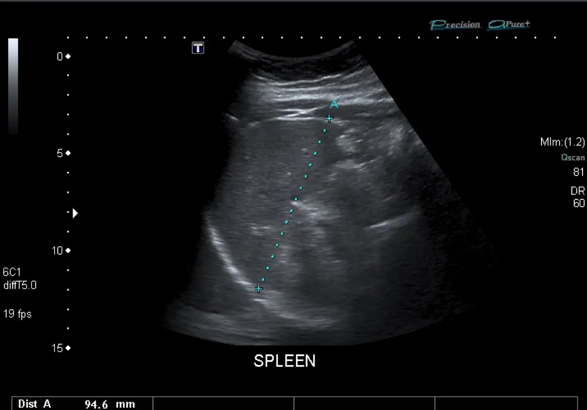
If your dog's abdominal ultrasound report mentions a "splenic mass", a "splenic nodule", or a "lesion on the spleen", it is completely understandable to feel a jolt of fear. The most important thing to know up front is that this finding is not an automatic diagnosis of cancer, and it is not an automatic death sentence. Many splenic masses turn out to be benign, and even when they are not, dogs can do well with the right plan. Here is what the finding actually means, the benign possibilities, the one situation that is a genuine emergency, and the steps that lead to a clear answer.
What a splenic mass or nodule means
The spleen is a soft organ that sits in the left side of the belly. It filters blood, stores red blood cells, and helps the immune system. Because it is packed with blood vessels, it is a common place for lumps to form as a dog ages. A "nodule" simply means a small lump; a "mass" usually means a larger one. Neither word tells you, on its own, whether the growth is harmless or serious.
On ultrasound, your veterinarian can see the size and shape of the lump, whether it is solid or filled with fluid or blood, and whether there is any free fluid in the abdomen that might signal bleeding. That is a lot of useful information. What ultrasound cannot do is look at the individual cells, and that is the catch: the same grainy pattern on the screen can be a completely benign lump or an aggressive tumor.
- A nodule is a small growth; a mass is a larger one, but size alone does not equal danger
- Ultrasound shows structure and bleeding, not the actual cell type
- Splenic lumps become more common with age, especially in medium and large breeds
- Some are found by chance on a scan done for an unrelated reason
The double two-thirds rule of thumb, and its limits
You may come across a well-known teaching guideline called the "double two-thirds" rule. Historically it held that roughly two-thirds of splenic masses in dogs are malignant, and that of those malignant ones, roughly two-thirds are a blood-vessel tumor called hemangiosarcoma. It is a memorable phrase, and it has shaped how vets think for years.
But treat it as a rough rule of thumb, not certainty. Newer studies report figures that vary quite a bit, and the odds shift with the size of the mass, whether the dog is bleeding, the breed, and other details. Plenty of dogs fall outside the two-thirds pattern in both directions. Most importantly, no percentage applies to your individual dog until the tissue is actually examined. The rule is a reason to take the finding seriously and investigate, not a verdict.
Benign possibilities worth knowing about
A meaningful share of splenic masses are not cancer at all. Knowing the common benign causes can take some of the fear out of the waiting.
- Hematoma — a pocket of clotted blood that can grow surprisingly large and look alarming on ultrasound, yet is entirely benign
- Nodular hyperplasia — a harmless overgrowth of normal splenic tissue, very common in older dogs
- Splenic torsion — the spleen twists on its blood supply; it is not a tumor, but it is itself an urgent surgical problem, so it needs prompt veterinary care rather than a wait-and-see plan
- Other benign growths — such as a hemangioma, the non-cancerous cousin of hemangiosarcoma
The frustrating reality is that a benign hematoma and a malignant hemangiosarcoma can look almost identical on a screen. That visual overlap is the single biggest reason the imaging finding, on its own, cannot close the case.
The one real emergency to watch for
There is one scenario that turns a splenic mass into an emergency: rupture. Because these lumps are full of blood vessels, a mass can suddenly bleed into the abdomen, sometimes with little warning. This is the situation to know cold, because acting fast genuinely matters.
Call a veterinarian or emergency clinic immediately if your dog shows any of these signs:
- Sudden weakness, wobbliness, or collapse
- Pale or white gums instead of healthy pink
- A belly that looks swollen or distended
- Fast, heavy breathing or a racing heart
- Unusual lethargy that comes on quickly, sometimes after a brief rally
These signs point to internal bleeding, and they need urgent care rather than a wait-and-see approach. If your dog is bright, eating, and behaving normally, you have time to plan the workup calmly with your own vet, which is the far more common situation once a mass is found incidentally. It is worth reading up on other findings your vet may look for on the same abdominal study, and comparing notes with related pieces such as how other common canine diagnoses are worked up.
How the mass is worked up and diagnosed
Once a splenic mass is found and your dog is stable, the goal is to understand the whole picture before deciding on surgery. This process is called staging, and it usually includes a few steps.
- Chest imaging — X-rays or a CT scan of the chest to check whether anything has spread to the lungs
- Heart ultrasound (echocardiogram) — because hemangiosarcoma can also involve the heart, an echo is often recommended; if you are curious how heart scans work, our piece on echocardiography in pets explains the basics
- Bloodwork — to assess anemia, clotting, and overall health for anesthesia
- Abdominal CT — sometimes added for a more detailed map before surgery
Here is the part that surprises many owners: vets generally do not biopsy a suspected bleeding splenic mass with a needle beforehand, because that can provoke dangerous bleeding. Instead, the definitive step is usually to remove the entire spleen surgically, a procedure called splenectomy, and send it to a pathologist. Examining the tissue under a microscope, called histopathology, is the only way to know for certain whether the mass is benign or malignant, and exactly what type it is. Dogs live perfectly well without a spleen, so removal is both the diagnosis and, in benign cases, the cure.
Why a second read can help
A splenic mass sits at a genuine crossroads: the same ultrasound picture can point toward a benign hematoma or a serious tumor, and the plan that follows, from staging to surgery timing, hinges on details that are easy to weigh differently. Having a fresh expert look at the images can add real reassurance before you commit to a path. DocOrbit offers a veterinary second opinion on your pet's imaging that you can bring to your own vet or surgeon, so the surgical and oncology plan is built on a careful, unhurried reading. It does not replace your treating veterinarian; it gives you and your vet another informed set of eyes.
Is a splenic mass in a dog always cancer?
No. A splenic mass is not automatically cancer, and it is definitely not an automatic death sentence. A commonly cited rule of thumb suggests that around two-thirds of splenic masses in dogs turn out to be malignant, but newer studies vary and many masses are benign, such as a hematoma or nodular hyperplasia. An ultrasound alone cannot tell benign from malignant, so the finding is a reason to investigate calmly, not to give up hope.
What are the signs that a splenic mass has ruptured?
A ruptured splenic mass causes bleeding into the belly, and the classic signs come on suddenly: weakness or collapse, pale or white gums, fast breathing, a distended or painful abdomen, and a racing heartbeat. This is an emergency. If you see these signs, take your dog to a veterinarian or emergency clinic right away rather than waiting for a scheduled appointment.
Can an ultrasound tell if a splenic mass is benign or malignant?
No. Ultrasound is excellent at finding a mass, measuring it, and checking whether it has bled, but it cannot reliably tell a benign lump from a cancer just by how it looks. Benign hematomas and malignant tumors can appear almost identical on the screen. The only way to know for certain is to examine the tissue under a microscope after the spleen is removed.
What is the double two-thirds rule for splenic masses in dogs?
It is an old teaching rule of thumb: roughly two-thirds of canine splenic masses are malignant, and of those malignant ones, roughly two-thirds are a tumor called hemangiosarcoma. It is only a rough guide, not a diagnosis. Newer studies show the real numbers vary quite a bit, and plenty of dogs fall outside the rule, which is exactly why tissue testing is needed to be sure.
Why is a splenic mass not biopsied before surgery?
Because sticking a needle into a splenic mass that may be filled with blood can trigger serious bleeding. For that reason vets usually avoid blindly biopsying a suspected bleeding splenic mass. Instead, the standard approach is to stage the disease, then remove the whole spleen surgically and send it for histopathology, which gives both a treatment and a definitive diagnosis at the same time.
Key takeaways
- A splenic mass on ultrasound is not automatically cancer, and not a death sentence
- The double two-thirds rule is a rough guide only; imaging alone cannot diagnose the mass
- Benign causes include hematoma, nodular hyperplasia, and splenic torsion
- Sudden collapse, pale gums, or a distended belly can mean rupture and need emergency care
- Staging plus splenectomy with histopathology is the only way to a definitive answer
Can I get a second opinion on my pet's ultrasound online?
Yes. Online veterinary radiology second opinions run entirely from home: you upload your images, with no CD to post and no second trip, and a subspecialist veterinary radiologist re-reads the study and writes a structured report you can give to your own vet. DocOrbit returns that signed second read in 24 to 48 hours. You can then message the veterinary radiologist with questions about the ultrasound and about what the report actually means for your pet.
This article is for general information only and is not veterinary advice. Always discuss your animal's imaging results and next steps with a qualified veterinarian.