
If your dog has started to sway on its back legs, scuff its toenails or walk as if the floor were slippery, and your vet has said "wobbler syndrome" or "cervical spondylomyelopathy," it is natural to feel frightened. The reassuring part first: wobbler syndrome is a well-understood problem of the neck, it is not caused by anything you did, many dogs stay comfortable for years with the right treatment, and a neck MRI shows your vet exactly where the trouble is. Here is what the name means, which dogs get it, what the scan shows and how it is treated.
What "wobbler syndrome" means
Wobbler syndrome gets its name from the swaying, drunken walk it causes. Vets call it cervical spondylomyelopathy, or CSM for short: cervical means the neck, spondylo refers to the neck bones (vertebrae) and myelopathy means a problem of the spinal cord.
The spinal cord runs through a tunnel formed by the neck bones, carrying messages between the brain and the legs. In a wobbler dog that tunnel is too narrow in one or more spots, or something has grown into it, so the cord is squeezed. The nerve fibers that steer the back legs sit on the outside of the cord and tend to be pinched first, which is why the hind end usually wobbles before the front legs are affected.
Two forms, two kinds of dog
Vets recognize two main patterns, and they tend to affect different dogs.
- Disc-associated: the soft cushion (disc) between two neck bones bulges up into the tunnel, often together with a thickened ligament. This is the form of middle-aged and older large dogs, classically the Doberman Pinscher, and it most often involves the lower neck, between the fifth and sixth or the sixth and seventh neck bones (C5-C6 and C6-C7). Rottweilers, Weimaraners and other large breeds can also be affected.
- Bone-associated: the vertebrae are misshapen, or the small joints between them have overgrown, so the tunnel is narrowed. This is the form of young, fast-growing giant breeds, classically the Great Dane, and it often involves the upper and middle neck. Mastiffs and Borzois, among other giant breeds, are also reported.
Some dogs have a mix of both. The tendency is inherited, and in growing giant-breed puppies very rapid growth and overfeeding are suspected to add to the risk, which is why vets advise a large-breed puppy food and a lean, steady build. None of this means you did anything wrong.
Signs owners notice
Signs usually creep in over weeks or months, and at first they are easy to explain away as clumsiness, growing up or getting older.
- A swaying, loose-limbed walk in the back legs, worse on slippery floors or when turning
- Crossing the back feet, tripping, or scuffing the tops of the toenails so that they wear down
- A short, stiff, choppy stride in the front legs, as if the dog were walking on eggshells, which appears once the front end becomes involved
- A sore neck: reluctance to look up or down, a head held low, or a yelp when the head is moved
- In some dogs, thinning of the shoulder muscles
Some dogs, especially those with the disc-associated form, stay stable for a long time and then worsen suddenly when a disc bulges a little more into an already narrow tunnel.
A wobbly walk is not usually an emergency, and you should book a vet visit within a day or two if it is getting worse. But see a vet today if your dog suddenly cannot stand or walk, cries out when its neck is touched or seems to be struggling to breathe.
Several other problems can look like wobbler syndrome, which is why the vet's neurological exam comes first. A disc extrusion in the neck (IVDD) can cause neck pain and weak legs, big dogs with hip dysplasia often sway on their back legs, and a short stride in front can come from elbow dysplasia. Spinal tumors and degenerative myelopathy, a slow, usually painless spinal cord disease of older dogs, are other possibilities.
Why MRI is the test of choice
A plain X-ray shows bone and can hint at malformed neck bones, but it cannot show the spinal cord or the soft tissue pressing on it, and some dogs with odd-looking neck bones on X-ray have no signs at all. So an X-ray alone cannot tell your vet whether, where or how badly the cord is squeezed. MRI shows the cord, the discs and the ligaments directly, which makes it the usual test for planning treatment. CT, sometimes with a contrast dye injected around the cord (myelography), is a good alternative and shows the bones in fine detail.
Your dog needs a general anesthetic to lie still for the scan, and the team positions the neck gently because it can be unstable. The pictures are always read alongside your dog's neurological exam: the scan shows the anatomy, and the exam shows how the cord is actually working.
What the neck MRI shows
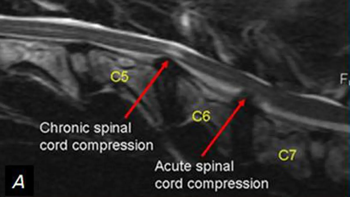
The picture at the top of this page is a side-on (sagittal) T2 MRI slice of a Doberman's neck, and the red arrows mark two places where the spinal cord is squeezed. On T2 pictures the fluid around the cord looks bright, so where that bright line thins or is pushed in, the tunnel is narrowed. The labels mark older (chronic) compression between C5 and C6 and recent (acute) compression between C6 and C7, and the uploader calls both levels "traction responsive," meaning the squeeze eased when the neck was gently pulled.
Radiologists look for:
- Where the tunnel is narrowed, level by level (named by the neck bones, such as C6-C7), and how many levels are involved, because many dogs have more than one
- What is squeezing the cord: a disc bulging up from below, a thickened ligament or overgrown joints from above and the sides, or a tunnel that is simply too narrow
- Which direction the pressure comes from, which helps decide the surgical approach
- Whether the cord itself looks injured: a bright patch inside the cord on T2 pictures points to swelling or scarring from the squeeze
- Whether the squeeze eases with traction (a light pull on the head), which some centers check with extra pictures and which helps decide between operations
Two honest caveats. A bright patch in the cord is a worrying sign, but it does not predict recovery on its own. And some dogs, Dobermans in particular, have disc bulges or narrowing on MRI without ever showing signs, so your vet matches the scan to the exam instead of treating the picture alone.
Treatment: rest and medication, or surgery
There are two broad routes, and the choice depends on how affected your dog is, which type of wobbler it has, how many levels are involved and your dog's age and general health.
Conservative (non-surgical) care suits dogs with mild signs or mainly neck pain. It means strictly limited activity for several weeks and then short lead walks, a harness instead of a collar, pain relief and anti-inflammatory drugs prescribed by your vet (sometimes a short course of steroids) and physiotherapy. Some dogs stay stable this way for a long time, but signs often return or progress, especially once normal activity resumes.
Surgery aims to take pressure off the cord and sometimes to hold the neck steady afterward. The operation is chosen to fit the cause: removing disc material through the underside of the neck (a ventral slot), opening the bone over the cord (a dorsal laminectomy), or holding the bones apart with a spacer and implants (distraction and stabilization). It is major surgery, best done by a surgeon or neurologist who does it regularly. Most dogs improve, though recovery takes weeks to months of rest and rehabilitation. Risks include anesthetic and implant problems, and a "domino" effect, where the neighboring disc starts to bulge months or years later.
Looking after a wobbler at home
- Use a harness instead of a collar, so a pull on the lead does not jerk the neck
- Put non-slip rugs on smooth floors, use a ramp for the car and the sofa, and block off stairs
- Keep exercise calm and on a lead, instead of rough play, ball chasing or jumping
- Keep your dog lean, because extra weight makes walking harder
- Never give human painkillers such as ibuprofen or acetaminophen (paracetamol), which can be toxic to dogs, and give only what your vet prescribes
What the outlook looks like
Wobbler syndrome is usually lifelong, so honest expectations help. Many dogs treated before they lose the ability to walk improve and stay comfortable for a long time, and surgery generally gives the better chance of lasting improvement in severely or progressively affected dogs. A perfectly normal walk is not guaranteed, and some dogs need further treatment over time.
Why a second read can help
A neck MRI is a stack of detailed slices, and many of the calls on it are judgments: which levels are really squeezed, whether a ligament or a disc is the main culprit, which direction the pressure comes from and whether a bright patch in the cord matters. Those details steer the choice between rest and major spinal surgery. Before you commit, you can ask a veterinary radiologist to re-read the MRI you already have.
DocOrbit offers a veterinary radiology second opinion online: a veterinary radiologist re-reads your dog's own MRI, and you can ask what the findings mean for treatment. A radiologist cannot examine your dog or feel how its legs respond, so this gives you and your vet another careful look at the same images and does not replace the vet or neurologist who treats your dog.
Is wobbler syndrome an emergency?
Usually not. A dog that is wobbly but still walking can be seen within a day or two, and many are managed over time. It becomes an emergency if your dog suddenly cannot stand or walk, cries out in pain or struggles to breathe, so call a vet right away if that happens.
Is wobbler syndrome painful for my dog?
It can be. Many dogs, especially those with the disc-associated form, have a sore neck and may yelp or hold the head low, while others are wobbly without obvious pain. If your dog seems uncomfortable, ask your vet about pain relief rather than giving human medicines, which can be dangerous.
Can a dog with wobbler syndrome have a good quality of life?
Many can. With sensible management or surgery, plenty of dogs stay comfortable and active for years, although the condition is usually lifelong and can progress or need further treatment. Your vet or a neurologist can say what is realistic for your dog.
Key takeaways
- Wobbler syndrome (cervical spondylomyelopathy) is a narrowing of the spinal canal in the neck that squeezes the spinal cord and causes a swaying walk, usually in the back legs first
- The disc-associated form affects middle-aged and older large dogs such as Dobermans, and the bone-associated form affects young giant breeds such as Great Danes
- MRI shows the cord and the cause of the squeeze, which an X-ray cannot, and is always matched to the neurological exam
- Mild cases may be managed with restricted activity, a harness and medication, while severe or worsening cases often need surgery
- A dog that suddenly cannot stand or walk needs a vet today
Can I ask a veterinary radiologist about my dog's MRI report online?
Yes. Online veterinary radiology second opinions run entirely from home: you upload your dog's images, with no CD to post and no second trip to a specialty clinic, and a subspecialist veterinary radiologist re-reads the study and writes a structured report you can give to your own vet. DocOrbit returns that signed second read in 24 to 48 hours, and you can message the veterinary radiologist afterward with questions about the MRI and about what it means for your dog.
This article is for general information only and is not veterinary advice. Always discuss your animal's imaging results and next steps with a qualified veterinarian.